In vitro Acoustic Radiation Force Impulse Imaging of Cardiac Ablation Lesions
Shruti Agashe, Stephen Hsu, Gregg Trahey, Patrick Wolf
Duke University, Department of Biomedical Engineering, Durham, NC, 27708
Abstract
Radio frequency ablation (RFA) lesions of cardiac tissue can not be visualized in vivo using any commonly used imaging technology. Lesion to lesion continuity and depth are both important for determining the successful outcome to RFA procedures. An in vitro chamber for making accurate geometric measurements of RF ablation lesions was designed and constructed. Acoustic radiation force impulse (ARFI) imaging was used to characterize lesions created in the chamber by RF ablation of ovine ventricular tissue. The boundaries of the lesions and the separation between lesions were imaged using ARFI and subsequently determined from histological sections. The ARFI imaging prediction of the lateral boundaries of the lesion correlated well with the actual lesion boundaries. The depth estimation from ARFI however was not as accurate. These results show the promise for using ARFI imaging in vivo during RF ablation for assessing the lesion size and continuity and ensuring complete blockage of the abnormal conduction pathways in atrial fibrillation and other cardiac arrhythmias
Introduction
Cardiac ablation has become the preferred treatment for various cardiac arrhythmias [2]. The ablation process uses a catheter based procedure and RF energy to coagulate an area of the myocardium and destroy the abnormal electrical pathway within the heart. For the procedure to be successful, the lesion must be large enough such that the abnormal pathway is blocked completely. It therefore follows that an imaging modality that is capable of visualizing the exact extent of the lesion would be an important asset to the ablation process [3]. However, the current clinically used methods of echocardiography(ICE) cannot differentiate normal and ablated myocardium. Acoustic radiation force impulse (ARFI) imaging uses the difference in the viscoelastic properties between the ablated and normal tissue to form an image of the lesion. Therefore cardiac ARFI can potentially directly visualize the development of the lesion as the ablation procedure progresses using the property of the changing myocardial stiffness [4]. The existing technology uses catheter based intracardiac anatomical mapping to guide the placement of the lesion. It is expected that ARFI imaging of the lesion would help predict the exact dimensions of the lesions and identify any incomplete lesion areas in real-time[1]. We designed and tested a system which would mimic the in vivo ablation procedure. This setup was then used to characterize the lesion using ARFI and direct comparisons between ARFI imaging and actual lesion size were made.
Methods
Image

| Figure 1) The photograph shows a chamber that was designed to mimic in-vivo cardiac ablation procedures. A Plexiglas® chamber containing a mixture of 0.9% saline and deionized water was used for the study. This chamber had an outlet and an inlet from a pump which continuously maintained a flow in the reservoir. A sheet of Aluminum foil (8cm X 8cm) ,placed in the saline solution approximately 15cm behind the piece of tissue and taped to the rear face of the box, acted as the neutral electrode in all of the ablation procedures. At the center of the box a vertical mount was designed to hold sound absorbing backing material behind the section of myocardium. |
Image

Figure 2) A Cardiac Pathways (Sunnyvale,CA) RF ablation device with a Boston Scientific (Natick, MA) SteeroCath catheter was used to create lesions in the myocardium. A guide sheath with a valve was used to introduce an 8 French ablation catheter from the front face of the box. An AcuNav{TM}, intra-cardiac probe was mounted on a translational and rotational stage and introduced into the saline solution. This stage was designed to preserve all six degrees of freedom and with no relative movement between the probe and the guide from the rotational stage. The 10 French, 64 element, 7 mm aperture diagnostic ultrasound catheter was attached to the Siemens SONOLINE Antares{TM} ultrasound scanner to create conventional B-mode and ARFI images. All B-mode images and ARFI sequences were acquired with a center frequency of 7.27 MHz and a pulse repetition frequency of 16.2 kHz . | |
Image

| Figure 3) The tissue holder had two slits cut into its horizontal faces at angles of 30 degrees and 60 degrees as shown. |
Procedure and Results
Ovine hearts were obtained from Hyclone labs and were frozen. A block of heart tissue (either from the left or right ventricle) cut to an appropriate size was thawed and then degassed. The tissue was placed in the designed tissue box (figure 2) with the endocardial surface exposed to the saline solution. A pulsatile flow was maintained around the tissue using a pump. The ablation catheter was introduced from the front of the box such that the tip was touching the tissue with adequate pressure to generate a sizeable lesion. Two experimental protocols were followed for characterizing the lesion.
Part A
This part of the experiment was to characterize the lesion size and shape and to measure the separation between lesions in a tissue slice. The AcuNav was first translated to image the tissue on one of the slits on the front face. The probe was then rotated by an angle so that so that the tissue was imaged in the plane of the slit. The imaging angle was verified by observing the bright reflections within the B-mode image with needles placed through the top and bottom portions of the slit. A 'before' ARFI image (Figure 3a left) was acquired first. Next, the ablation catheter was pressed on the tissue block and a lesion was created using RF generator settings of 25 Watts and 30 seconds. After ablating the tissue, the catheter was retracted and a second ARFI image (Figure3a right ) was acquired in the same plane. The tissue was then removed from the tank and sliced using the slits as guides. This slice was then photographed and the digitized image was compared with the ARFI image to estimate lateral boundaries, depth and area of the lesion (Figure 3b).
Image Image

| Image Image

|
| Figure 3a) Shows ARFI images overlaid on the B-mode image. The image on the left is captured before ablating the tissue. The image on the right is captured after ablating the tissue and retracting the ablation catheter. | |
Image Image

|
| Figure 3b) Shows a photograph of the lesion (left) and the corresponding ARFI image (on the right) from 3a) The images have been shown on the same scale for comparison. |
For a second piece of tissue, two lesions were created along a vertical line (Figure 4a). These lesions were created with ablation settings of 28 Watts and 30 Watts for 30 seconds each. The same protocol as described above was followed. Only this time the AcuNav was translated vertically in line with the slit and images were captured to estimate the gap between the lesions. One of these images for the estimation of separation is shown in Figure 4b.
Image Image

| Figure 4a) Shows a photograph of the tissue slice cut along one of the slits (imaging plane) with the cross sections of the two lesions. | |
Image

| Image

| |
| Figure 4b) Shows ARFI images overlaid on the B-mode image. The image on the left is captured before ablating the tissue. The image on the right ('After' image) is captured after making two lesions separated by some distance vertically in the setup. | ||
Image Image

| Image Image

| ||
| Figure 4c) Shows a comparison between the lesion separation in the 'After' image and the digitized slice of tissue. The image on the bottom left is shown with the 'time to peak' parameter as an indication of stiffness of myocardium. The images have been shown on the same scale for comparison. | |||
Part B
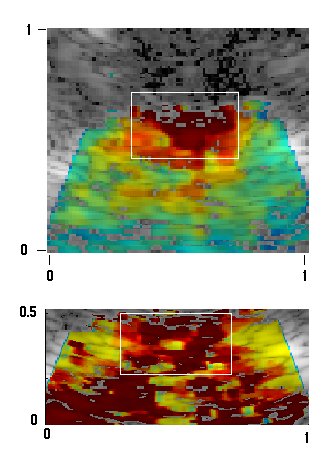
A second protocol was followed to estimate the surface area of the lesion. In this protocol, the ARFI probe was aligned to image the front face of the tissue. A lesion was created with RF generator settings of 25 Watts and 60 seconds. The imaging probe was positioned directly above the lesion and an image taken at this position was recorded as 0. The stage was translated in elevation and ARFI images were captured at every 0.4mm. The images were acquired until the lesion was no longer visible in the ARFI image. These set of images were processed to generate a C scan of the myocardial surface. The ARFI images with a surface contour estimation of the lesion are shown in figure 5)
Image Image

| Image Image

|
| Figure 5) Shows a reconstruction of the surface contour of the lesion obtained from a set of 2D ARFI images on the left and a photograph of the surface of the tissue with the lesion on the same scale to the right. | |
Discussion
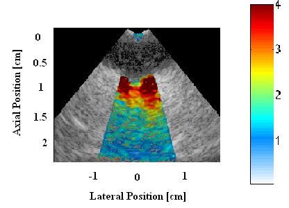
ARFI images of the lesion show smaller displacements in the areas containing the lesion. The ablated area therefore is stiffer tissue and shows lesser displacements in ARFI. Figure 3 shows a comparison between the photographed lesion and the ARFI image. The lateral extent and depth of the lesion predicted by ARFI is 0.4mm and 0.2mm while from the photograph the values are 0.5mm and 0.4mm respectively. In figure 4 a comparison between the gap estimation by ARFI and the digitized image is made. ARFI and the digital image estimate the separation to be 0.5mm. Also shown in figure 4c) is the "time to peak" ARFI image which also gives an estimate of the separation to be 0.5mm. We believe that other parameters may be capable of making better estimations of the depth independent of attenuation effects. A reconstruction of the surface contour of the lesion has been made as shown in figure 5. In our setup there are human errors as well as alignment errors which could be minimized in further experiments. All image segmentations have been done by hand and are subjective.
Conclusion
Our studies prove that ARFI techniques could be used to image the boundaries of the ablated area. However our results show better lateral resolution than depth resolution. From our results, it is further evident that ARFI can estimate the separation between lesions. This setup can be used for 3D imaging studies to compare ARFI estimations with the lesion, reconstructed using histology, to provide further proof in support of ARFI estimation of ablation. This apparatus is capable of giving direct quantitative comparisons between ARFI estimation and actual size of ablation lesions.
References
[1] Fahey BJ, Nightingale KR, McAleavey SA, Palmeri ML, Wolf PD, Trahey GE. "Acoustic radiation force impulse imaging of myocardial radiofrequency ablation: initial in vivo results." IEEE Trans. Ultrasonics Ferroelectrics and Frequency Control. 2005; 52(4): 631-641
[2] Stephen J Hsu, Stephen Smith, Julia Hubert and Gregg Trahey, "Ex Vivo Acoustic Radiation Force Impulse Imaging of an Ovine Heart Model"
[3] Stephen J Hsu, Richard Bouchard, Douglas Dumont, Patrick Wolf and Gregg Trahey, "In Vivo assessment of myocardial stiffness with acoustic radiation force impulse imaging"
[4] Stephen J Hsu, Brian Fahey, Douglas Dumont, Patrick Wolf and Gregg Trahey, "Challenges and Implementation of Radiation-Force Imaging with an Intracardiac Ultrasound Transducer"
Acknowledgments
This research was funded by NIH Grant #: R21-EB-007741. We would like to thank Siemens Medical Solutions USA, Inc. for their hardware and system support. We would like to thank Mr. Steven J Owen (Mechanical department-Duke University) for setting up the imaging chamber and his valuable suggestions in the course of designing the assembly.